
Figure 1.
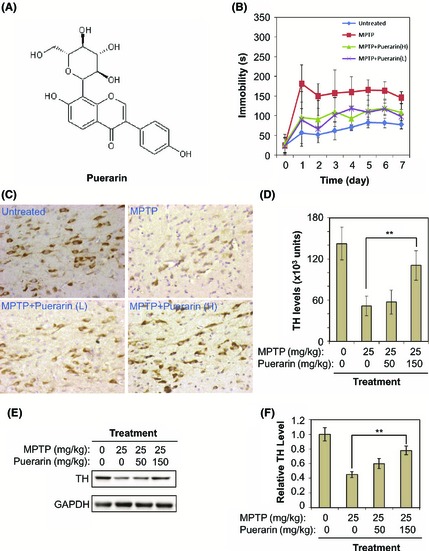
In vivo neuroprotective and neurotrophic activities of puerarin against MPTP neurotoxicity in a murine model of PD. (A) Chemical structure of puerarin. The structure was generated by ChemSketch software (http://www.acdlabs.com). (B) Amelioration of MPTP‐induced behavioral impairments. A total of 24 mice were divided into four groups (n = 6): Untreated, no treatment; MPTP, 25 mg/kg MPTP (i.p.) per day; MPTP+Puerarin (L), 50 mg/kg of puerarin (i.p.) and 25 mg/kg MPTP (i.p.) per day; MPTP+Puerarin (H), 150 mg/kg of puerarin (i.p.) and 25 mg/kg MPTP (i.p.) per day. The immobility of mice in forced swim test was recorded as an indicator of behavioral performance. (C) Reduction of MPTP neurotoxicity on tyrosine hydroxylase positive (TH +) dopaminergic neurons. The animals were treated as described in Panel B. TH protein was immunostained with specific antibody and visualized as dark brown stains produced by the HRP conjugate of secondary antibody. (D) Data analysis of TH immunostaining. Following detection described in Panel C, TH expression levels were determined by reading the optical density of stains. The mean values of the optical intensity for each group of animals (n = 6) were analyzed by one‐way ANOVA (**P < 0.01, Puerarin + MPTP group vs MPTP group). (E) Recovery of MPTP‐induced loss of TH expression. The animals were treated as described in Panel B. TH protein was determined by Western blotting with specific antibody. (F) Data analysis of TH expression. The optical density of TH bands in Panel C was determined by software Quantity One (Bio‐Rad). The mean values of the optical intensity for each group of animals (n = 6) were analyzed by Student's t‐test (**P < 0.01, Puerarin + MPTP group vs MPTP group).