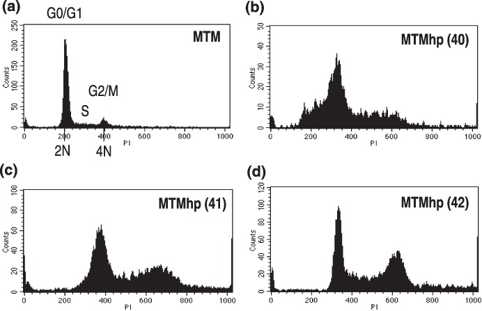
Figure 3.

(a) Primary mesothelial cells and spontaneously transformed mesothelial cell lines. (b) MTMhp (40), (c) MTMhp (41), and (d) MTMhp (42) were stained with propidium iodide and their DNA content assessed by flow cytometry. Each histogram plot is representative of data generated from at least three individual experiments.
