Figure 2.

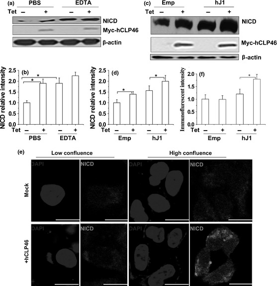
Overexpression of hCLP 46 enhanced Notch activation. (a) 293TRex‐hCLP46 cells were incubated for 48 h in absence (−) or presence (+) of 0.5 μg/ml Tet. Cells were treated with 5 mm EDTA or PBS and then cultured for additional 30 min. (c) 293TRex‐hCLP46 cells were incubated for 42 h in absence or presence of Tet and then co‐cultured for 5 h with 293T cells transfected with human Jagged1 (hJ1) or empty vector (Emp) for 36 h. Immunoblot band intensities were quantified using loading controls (b, d). (e) 293TRex‐hCLP46 cells were seeded at low confluence (low) or high confluence (high) and were incubated for 48 h in absence or presence of Tet. Green fluorescence indicates that NICD and nuclei were counterstained with DAPI (blue). Confocal images are representative of results obtained in three separate sets of experiments. Bar = 20 μm. Immunofluorescence intensities were quantified (f) and data represent mean ± SD of three independent experiments. *P ≤ 0.05.
