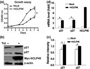
Figure 4.

hCLP 46 inhibited cell proliferation and increased CDKIs p21 and p27. (a) 293Trex‐hCLP46 cells were incubated in absence or presence of 0.5 μg/ml Tet. At indicated time points, cells were harvested and incubated with MTT; absorbance at 492 nm was determined. All time points were assayed in triplicate. Cells were incubated for 48 h in absence or presence of Tet (b) and cell lysates were immunoblotted with indicated antibodies. (c) Immunoblot band intensities were quantified using loading controls. (d) Total RNA was subjected to RT‐PCR analysis for p21, p27 and hCLP46. Data represent mean ± SD of three independent experiments. *P ≤ 0.05.
