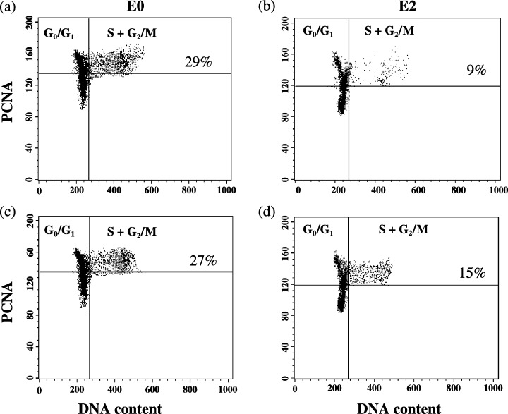
Figure 1.

Examples of T‐cell proliferation of two HTx recipients before (EO, parts a and c) and two hours (E2, parts b and d) after dosing of cyclosporin. Two colour flow cytometric analysis of PCNA expression and DNA content of two HTx recipients before (E0, parts a and c) and two hours (E2, parts c and d), after intake of the morning dose of cyclosporin. Dot plots show Con A stimulated lymphocytes in whole blood after three days incubation.
