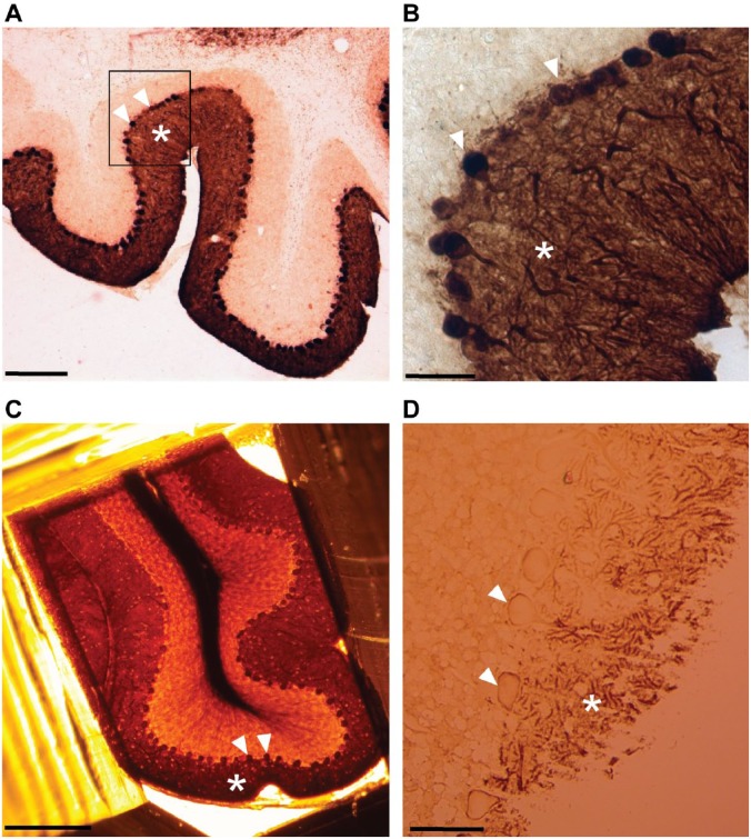
Figure 1.
Light microscopy imaging of Purkinje cells labeled with calbindin-D28k antibody. (A) Sagittal vibratome sections (80 µm) of cerebellum obtained after labeling with calbindin followed by DAB/GSSP reactions display characteristic dark brown staining patterns corresponding to bodies of stained neurons bodies (arrows) and dendrite area (asterisks) in the cortex of cerebellum. (B) The same section (A, black box) at a higher magnification showing calbindin staining in bodies of Purkinje cells (arrows) and in the dendritic area (asterisk). (C) Purkinje cell somata (arrows) and dendrite area (asterisk) visualized in a resin block after embedding for electron microscopy. (D) Visualization of labeled Purkinje cell neuropil (asterisk) and bodies (arrows) in a semithick section (0.5 µm). Scale bar, A = 500 µm, B, D = 50 µm, C = 250 µm. Abbreviations: DAB, diaminobenzidine, GSSP, gold-substituted silver peroxidase.

