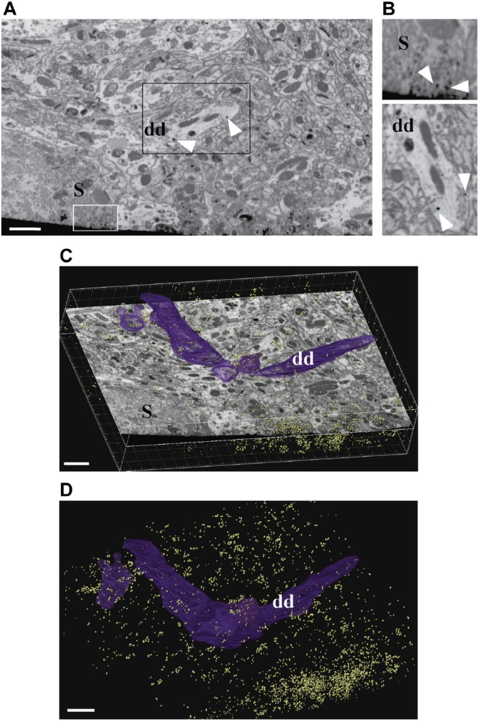
Figure 3.
FIB-SEM imaging of calbindin GSSP-labeled Purkinje cells. (A-B) A single block-face image from an FIB-imaged stack, showing a fragment of the Purkinje cell layer (S, Purkinje cell soma) with GSSP-positive areas in somata of Purkinje cells (white box) and in dendrites (dds; black box). (B) Higher magnification images of areas highlighted in 3A. Top, fragment of a body of a Purkinje cell showing calbindin immunolabeling as distinct round deposits that are darker than any elements of cellular ultrastructure, white arrowheads. Bottom, fragment of a dendrite of a Purkinje cell, rotated image. Note the highly contrasted, regularly shaped deposits corresponding to calbindin immunoreactivity. (C-D) The size and density of GSSP particles allows to easily follow and segment immunopositive structures. Individual GSSP particles shown in yellow and manual segmentation of a Purkinje cell dendritic segment in purple (dd). Dendrite indicates the same dendrite in 3A, 3B (bottom), 3C, and 3D. Imaging was performed using a ZEISS Auriga Crossbeam FIB-SEM. Scale bar = 200 µm. Abbreviations: FIB-SEM, focused ion beam–scanning electron microscopy; GSSP, gold-substituted silver peroxidase.

