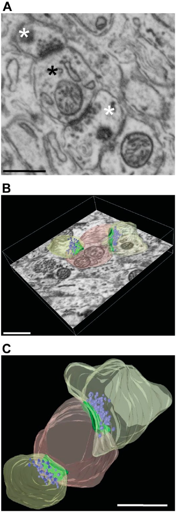
Figure 4.

FIB-SEM imaging of Purkinje cells synapses. (A) Details of synaptic ultrastructure after FIB-SEM imaging of an unlabeled area of a calbindin-stained specimen. A sample image of a postsynaptic bouton (black asterisk) making contact with two presynaptic elements (white asterisks), showing good preservation of ultrastructure. (B-C) Same synapse manually segmented and reconstructed; postsynapse is shown in pink, presynaptic profiles in yellow, AZ-PSD complex in green, and vesicles in purple. Imaging was performed using a FEI Scios DualBeam system. Scale bar = 100 µm. Abbreviations: FIB-SEM, focused ion beam–scanning electron microscopy; AZ, active zone; PSD, postsynaptic densities.
