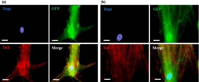
Figure 5.

Cardiomyogenic differentiation of expanded cardiac atrial appendage stem cells. Immunofluorescence images illustrating cTnT (red, a) and cTnI staining (red, b) on GFP+ CASCs (green; P4–P9) after 1 week in co‐culture with NRCMs. Nuclei stained with 4′,6′‐diamidino‐2‐phenylindole (DAPI) (blue). N ≥ 3 measurements at each passage. Scale bar = 20 μm.
