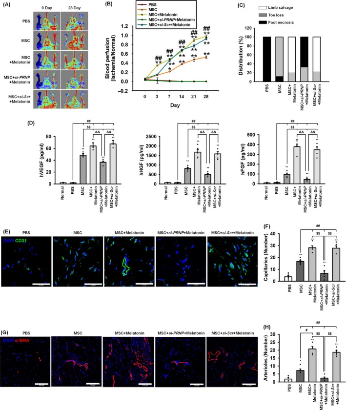
Figure 7.

Assessment of functional recovery and neovascularization in a murine hindlimb ischaemia model. A, Blood perfusion was assessed using laser Doppler perfusion imaging analysis in the ischaemic limbs of mice injected with PBS, MSCs (MSC), melatonin‐treated MSCs (MSC + Melatonin), MSCs treated with melatonin and PRNP siRNA (MSC + si‐PRNP + Melatonin), or MSCs treated with melatonin and scrambled siRNA (MSC + Scr‐siRNA + Melatonin). B, The blood perfusion ratio was measured by LPDI analysis (blood flow in the left ischaemic limb/blood flow in the right non‐ischaemic limb). Values represent the mean ± SEM. *P < 0.05; **P < 0.01 vs PBS, and ## P < 0.01 vs MSC. C, Distribution of the different outcomes (limb salvage, toe loss and foot necrosis) in each of the treatment groups at post‐operative day 28. D, Secretion of human VEGF, HGF and FGF from the ischaemic‐injured tissues in each group was assessed by ELISA. Values represent the mean ± SEM. **P < 0.01 vs normal, ## P < 0.01 vs PBS, $$ P < 0.01 vs MSC, and && P < 0.01 vs MSC + si‐PRNP + Melatonin. E‐G At post‐operative day 28, the formation of capillaries and arterioles were analysed using immunofluorescent staining for CD31 (green; scale bar = 50 μm; E) and α‐SMA (red; scale bar = 100 μm; G). The capillary (F) and arteriole (H) densities were quantified as the number of CD31‐positive cells and α‐SMA‐positive cells, respectively. Values represent the mean ± SEM. *P < 0.05; **P < 0.01 vs PBS, # P < 0.05; ## P < 0.01 vs MSC, $$ P < 0.01 vs MSC+si‐PRNP + Melatonin
