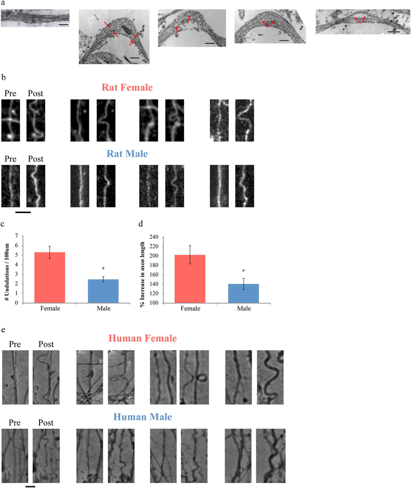
Fig. 5.
Dynamic stretch-injury of axon tracts in vitro. (a) TEM images of axons pre- and post-injury show change of morphology from straight to undulated due to mechanical breaking of microtubules (red arrows depict break sites) (Scale bar, 200 nm). (b) Representative images showing injured female rat axons with more extensive and larger undulation formation compared to male axons (Scale bar - 5 μm), (c, d) Undulations were analyzed with respect to the number of undulations/100 μm (c) and percent increase in axon length (d). (e) The same sex difference was observed for injured human axons (Scale bar - 5 μm). Data is shown as mean ± s.e.m. (d = 6, e = 7 independent experiments). Statistical significance (* p < 0.01) was evaluated by Students t-test. (For interpretation of the references to color in this figure legend, the reader is referred to the web version of this article.)

