Figure 1.

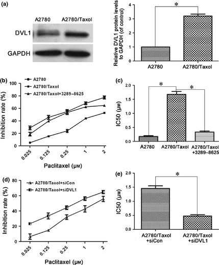
Silencing DVL1 enhanced the sensitivity of A2780/Taxol ovarian cancer cells to paclitaxel. (a) Western blotting of DVL1 expression in A2780 and A2780/Taxol cells. GAPDH was used as the internal control. The left panel shows representative Western blots, and the right panel shows quantitation of the Western blots to show the relative DVL1 expression normalized to GAPDH control. (b) The drug sensitivity of A2780, A2780/Taxol and compound 3289‐8625‐treated A2780/Taxol cells was assessed using MTT assays to measure cell viability. (c) The IC50 of paclitaxel in A2780, A2780/Taxol and compound 3289‐8625‐treated A2780/Taxol cells. The IC50 of paclitaxel was calculated from the survival curves generated in (b) using the Bliss method. (d) The drug sensitivity of A2780/Taxol cells transfected with siDVL1 or siControl. (e) The IC50 of paclitaxel in transfected A2780/Taxol cells, which was calculated from the survival curves in (d) using the Bliss method. Each experiment was performed in triplicate. *P < 0.05.
