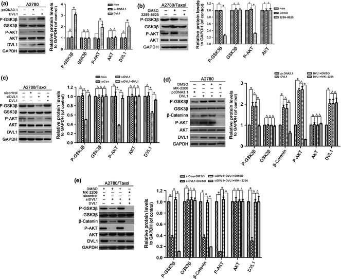
Figure 4.

The effect of DVL1 on AKT/GSK‐3β/β‐catenin signalling. Western blotting for phospho‐GSK‐3β (Ser‐9), GSK‐3β, phospho‐AKT (Ser‐473), AKT and DVL1 (when pcDNA3.1‐DVL1‐ or siDVL‐transfected cells were included) in (a) A2780 cells transfected with pcDNA3.1‐DVL1 or pcDNA3.1 for 72 h, (b) A2780/Taxol cells treated with or without compound 3289‐8625 for 72 h, (c) A2780/Taxol cells transfected with siControl, siDVL1 or siDVL1 plus pcDNA3.1‐DVL1 for 72 h, (d) A2780 cells transfected with pcDNA3.1 or pcDNA3.1‐DVL1 for 72 h, and then treated with the AKT inhibitor MK‐2206 for 5 h, and (e) A2780/Taxol cells transfected with siControl, siDVL1 or siDVL1 plus pcDNA3.1‐DVL1 for 72 h, and then treated with MK‐2206 for 5 h. GAPDH was used as the internal control. Left panels show representative Western blots, and right panels show the relative quantification after normalization to GAPDH. Each experiment was performed in triplicate. Δ P > 0.05; *P < 0.05.
