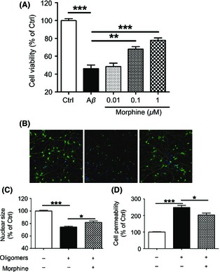
Figure 1.

Morphine attenuated Aβ oligomers‐induced neurotoxicity in primary cultured cortical neurons. (A) Primary cultured rat cerebral cortical neurons were incubated with 0, 0.01, 0.1, and 1 μM morphine for 2 h before 1 μM Aβ‐(1‐40) oligomers was added. Cells were then cultured for 24 h, and cell viability was measured by CCK‐8 assay. (B) Primary cultured rat cerebral cortical neurons were incubated with 1 μM morphine for 2 h before 1 μM Aβ‐(1‐40) oligomers was added for another 24 h. Then, the neurons were stained with Hochest 33342 for nuclei and neurite primary antibody for cell bodies and neurites. Representative images were taken by Cellomics KineticScan HCS Reader. Magnification was 512 × 512 pixels (10 × for parameter settings, one pixel represents 0.625 μM). (C–D) Primary cultured rat cerebral cortical neurons were incubated with 1 μM morphine for 2 h before 1 μM Aβ‐(1‐40) oligomers was added for another 24 h. Nuclear size (C) and cell permeability (D) were measured and calculated using multiparameter HCS cytotoxicity kit and Cellomics BioApplication Modules. *P < 0.05; **P < 0.01; ***P < 0.001.
