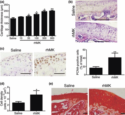
Figure 3.

rhMK stimulated proliferation of articular chondrocytes in mice after systemic delivery. Mice received a daily subcutaneous injection of vehicle saline or rhMK at different dosages for 7 days. After 15 days from first injection, the mice were killed. Histological examination and cell analysis of articular cartilage and chondrocyte were performed as described in the Materials and methods section. (a) Tibial plateau thickness of articular cartilage of three animals per treatment group is shown. rhMK stimulated enlargement of tibial plateau cartilage in a dose‐dependent manner. (b) Representative tibial cartilage tissue sections from mice treated with saline or rhMK (300 μg/kg). HE staining, bar = 200 μm. (c) Cell proliferation marker PCNA expression analysis in condylar cartilage tissue sections from the mice treated with saline or rhMK (300 μg/kg). Left, representative tissue sections immunohistochemically stained for PCNA, bar = 50 μm; right, quantitative and statistical analysis of PCNA‐positive chondrocytes (n = 3). (d) Chondrocyte density is shown for tibial cartilage from mice treated with saline or rhMK (300 μg/kg) (n = 3). (e) Condylar cartilage sections from mice treated with saline or rhMK (300 μg/kg) were stained with cartilage matrix‐specific dye Safranin O (bar = 50 μm). Similar intensity of staining is shown. All data are presented as mean ± SD. Three independent experiments showed similar results. Values were compared to the saline group using the two‐tailed Student’s t‐test. *P < 0.05, **P < 0.01.
