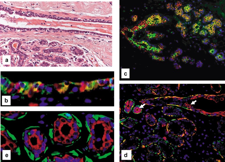
Figure 1.

Normal breast epithelium. (a) Small duct and adjacent lobule. Note that the normal epithelium consists of a bilayer with luminal glandular and basal myoepithelial cells (haematoxylin and eosin). (b) Double‐fluorescence staining of the epithelium of a small duct for CK5 (FITC, green) and CK8/18/19 (Cy3, red) displaying few CK5+ progenitor cells (green signal), intermediary cells (hybrid signal) and CK8/18+ glandular cells (red signal). (c) Double‐fluorescence staining of the epithelium of two terminal ducts and adjacent lobules of a resting breast for CK5 (green signal for FITC) and CK8/18 (Cy3, red) revealing the same cellular components as seen in the small duct in (b), indicating a relatively immature glandular epithelium. (d) Double‐fluorescence staining of the epithelium of a small duct and adjacent lobule of a resting breast for CK5 (FITC, green) and SMA (Cy3, red). The arrow marks a progenitor cell expressing CK5 alone. Note that most cells in the outer layer represent intermediary myoepithelial cells expressing both CK5 and SMA (hybrid colour). The differentiated myoepithelial express only SMA (green signal). Note also that the inner epithelium of both duct and of two acini for CK5 (FITC, green) and SMA (Cy3, red) contain precursor cells. (e) Double‐fluorescence staining of the epithelium of lobular acini (red signal) Panels for SMA (FITC, green) and CK8/18 (Cy3, red) clearly showing a glandular and myoepithelial cell lineage. Note the absence of any transitional cells, indicating that there is no transdifferentiation between those two lineages.
