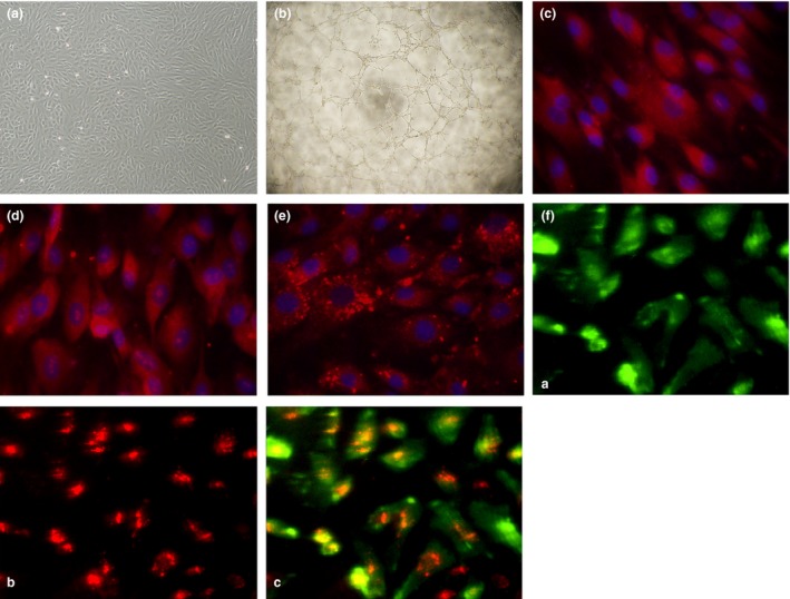
Figure 1.

(a) Typical morphological appearance of EPC s in culture (10× objective lens). Typical homogeneous and cobblestone‐like morphology of cells after 14 days culture. (b) Tube formation capacity on Matrigel matrix (10 × 4 objective lens). (c–f) Representative immunofluorescence staining of endothelial cell markers including stem cell marker CD34 (c, red), KDR (d, red), vWF (e, red). Cell nuclei were counterstained with DAPI in blue (40x). (f) The cultured cells were able to bind UEA‐1‐FITC (f‐(a), green) and take up DiI‐acLDL (f‐(b), red), (f‐(c), two‐colour overlay)
