
Figure 1.
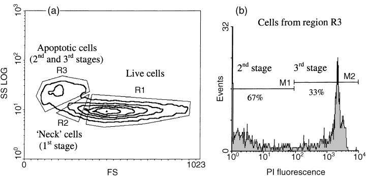
Different stages of apoptosis detected by flow cytometry in in vitro culture of EL‐4 cells. (a) Regions of live and apoptotic cells selected by their morphological features (forward and side light scatter) . (b) Second and third stages of apoptosis selected by the level of PI staining cells from apoptotic region identified by morphological features (region R3, second and third stages).