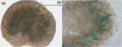
Figure 3.

Migration pattern of hAFSCs in developing embryonic kidney, histology of sectioned kidneys post‐injection of stem cells. (a) Lac‐Z staining confirming the presence of hAFSCs after 3 days in culture (×6). (b) Lac‐Z+ hAFSCs migrated to the periphery of the embryonic kidney after injection into the middle of organ (×8). hAFSCs, human amniotic fluid stem cells.
