
Figure 1.
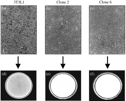
Differentiation of 3T3L1 cells into adipocytes upon hormonal induction and lack of differentiation in Raidd‐transfected 3T3L1 cells. (a, b, c) are photographs (100 × magnification) of cells 8 days after hormonal induction to differentiation. (a) shows parental 3T3L1 cells that differentiated normally after hormonal induction. In contrast, cells stably transfected with the murine Raidd cDNA did not undergo adipocyte differentiation in response to hormone treatment [(b) and (c) for clones 2 and 6, respectively]. (d, e, f) show cells stained with Oil Red O, which stains specifically the lipids. Dark staining is observed in the parental 3T3L1 cells (d) that accumulate triglycerides indicating appropriate differentiation into adipocytes. No staining is observed in the Raidd ‐transfected clones [(e, f), for clones 2 and 6, respectively] confirming the blockage of differentiation in these clones.