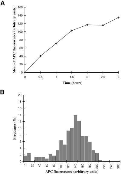
Figure 2.

Nuclear accumulation of APC‐NLS. (A) Nuclei from scrape‐ruptured quiescent cells were incubated for the indicated times in Xenopus egg extracts containing APC‐NLS. The APC fluorescence of individual nuclei was quantified by confocal microscopy and the mean fluorescence plotted as a function of time. (B) Frequency histogram of APC‐fluorescence for individual nuclei, for the 3 h time point in (A).
