
Figure 1.
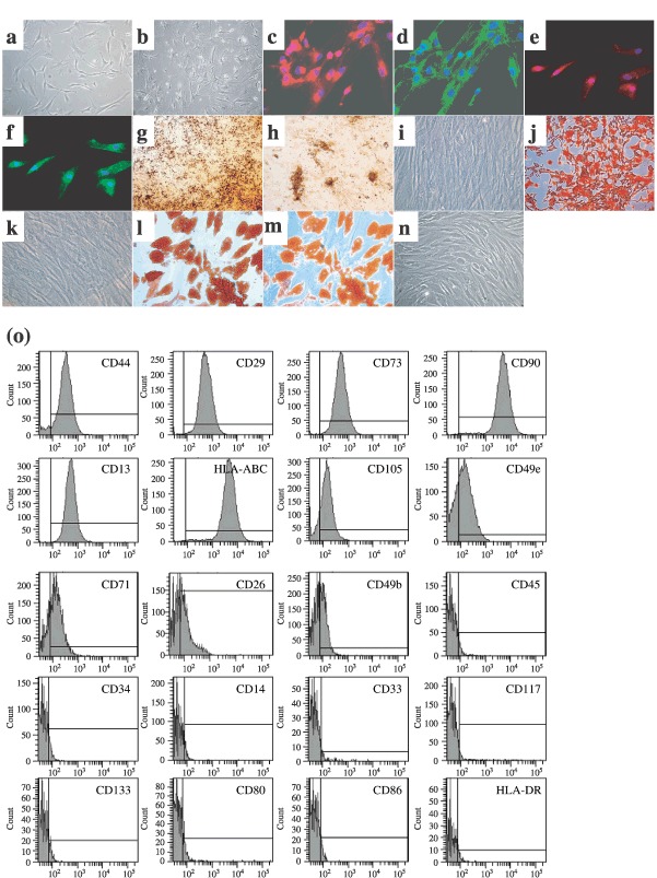
Characterization of MSCs. Example of MSCs morphology at passage 1, day 15 (a) that was altered at passage 8, day 99 (b). Expression of mesodermal antigens in MSCs at passage 2 as revealed by immunofluorescence [vimentin (c), fibronectin (d), ASMA (e), laminin (f), n = 3 each]. Staining of extracellular calcium matrix deposition by von Kossa (g, h) and alizarin red (j) colorations in MSCs after osseous differentiation. Accumulation of intracytoplasmic lipid‐rich droplets revealed by oil red O staining (l, m) after adipocyte differentiation of MSCs. Respective controls are provided with undifferentiated MSCs (i, k, n). Example of representative immunophenotype of MSCs at passage 2 (o). Analyzed epitopes are indicated in the histograms. Bars indicate the fluorescence level of corresponding isotype. Pictures magnifications are: ×100 (b, g), ×200 (j), ×300 (c–f) and ×400 (a, i, k–n).