
Figure 3.
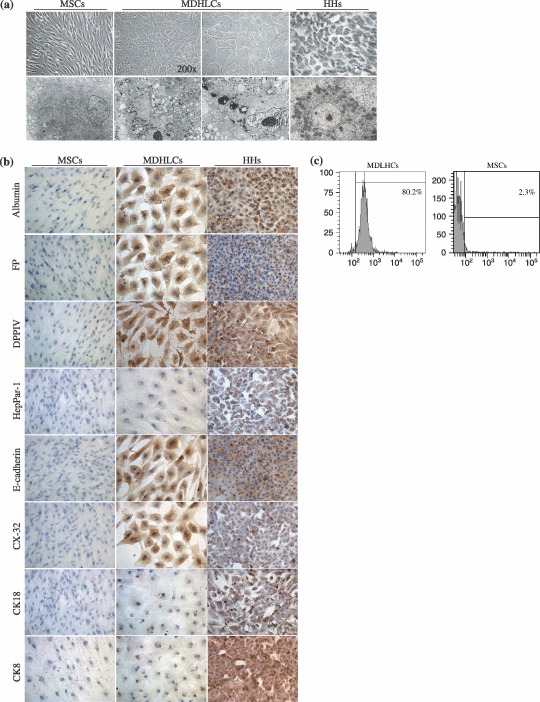
In vitro hepatocyte differentiation of human bone marrow‐MSCs. (a) Comparative aspect of MSCs, MDHLCs and HHs as observed in optic (OM) and electron (EM) microscopy. Pictures show that MDHLCs acquire round or polygonal‐shaped cells reduced in size and containing cytoplasmic granulations and central nucleus with prominent nucleolus. HHs comparative cell size was smaller. EM reveals that MDHLCs exhibited a higher content in cytoplasmic organites (endoplasmic reticulum, mitochondria) and a prominent nucleolus suggesting their enhanced metabolic activity. They produced intracytoplasmic glycogen vacuoles that are a hallmark of hepatocyte‐like functionality. Comparison with MSCs and HHs is provided. OM pictures were taken at magnification ×400 unless indicated and EM pictures magnification was ×4000. (b) MDHLCs immunostaining for hepatocyte markers showing positive expression of albumin, αFP, DPPIV and some epithelial markers (E‐cadherin, CX‐32) but not of HepPar‐1, CK8 and CK18. MSCs and HHs stainings were provided as negative and positive controls, respectively. Pictures were taken at magnification ×400. (c) Representative examples of albumin staining by flow cytometry in MDHLCs and MSCs. Bars indicate the fluorescence level of the corresponding isotype. αFP, α‐foetoprotein; CK18, cytokeratin 18; CX‐32, connexin‐32; DPPIV, dipeptidylpeptidase IV; HHs, human primary hepatocytes; MDHLCs, mesenchymal‐derived hepatocyte‐like cells; MSCs, mesenchymal stem cells.