
Figure 5.
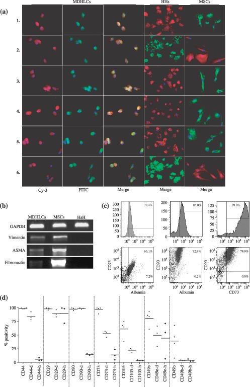
Characterization of the chimerical phenotype of MDHLCs. (a) Immunofluorescence assay showing co‐staining of MDHLCs, HHs and MSCs for albumin/fibronectin (1), ASMA/αFP (2), DPPIV/fibronectin (3), E‐cadherin/fibronectin (4), CX‐32/fibronectin (5) and vimentin/albumin (6). Pictures were taken at magnification ×300. (b) RT‐PCR analysis revealing the persistence of vimentin, ASMA and fibronectin expression in MDHLCs population. (c) Flow cytometry assay showing a representative example of albumin, CD73 and CD90 co‐staining on MDHLCs. (d) Comparative flow cytometric phenotype of MSCs (passage 2 to 4), MDHLCs (passage 1 to 3), and human hepatocytes. MDHLCs and hepatocytes are specified respectively by (‐d) and (‐h) suffixes. P values are indicated below graph (*P < 0.05, **P < 0.01, ***P < 0.001). αFP, α‐foetoprotein; ASMA, α‐smooth muscle actin; CX‐32, connexin‐32; DPPIV, dipeptidylpeptidase IV; HHs, human primary hepatocytes; MDHLCs, mesenchymal‐derived hepatocyte‐like cells; MSCs, mesenchymal stem cells.