
Figure 7.
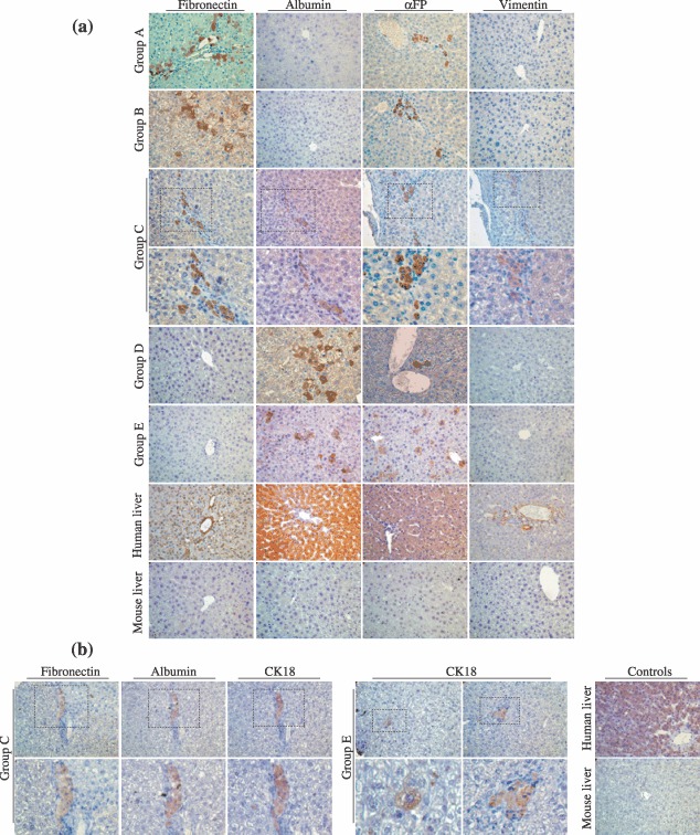
In vivo characterization of MSCs and MDHLCs after transplantation into SCID mice. (a) Analysis of in vivo differentiation potential of MSCs and MDHLCs. Pictures show immunostaining for human mesodermal (fibronectin, vimentin) and hepatocyte (albumin, αFP) antigens. In groups A and B, the engrafted cells formed clusters presenting staining for fibronectin or αFP separately. In group C, cell clusters co‐expressed mesodermal and hepatocyte markers as assessed by serial sections. In groups D and E, where MDHLCs were injected respectively into the spleen or the liver, the engrafted cell clusters expressed only hepatocyte markers. Stains were performed on human and mouse livers respectively as positive and negative controls. (b) Analysis of CK18 expression in groups C and E mice. In group C, the engrafted cells displayed co‐expression of fibronectin, albumin and CK18 whereas in group E, isolated staining for CK18 was observed in single or clustering cells. Control stains for CK18 were performed on human and mouse livers. Pictures were taken at magnification ×400. αFP, α‐foetoprotein.