
Figure 9.
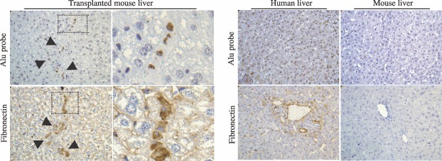
In vivo tracking of human cells. Pictures show that engrafted human cells were co‐stained at the nuclear level with Alu probe by in situ hybridization technique and at the cytoplasmic level with antihuman fibronectin antibody by immunohistochemical assay on serial sections. Human and mouse livers are provided respectively as positive and negative controls. Arrows indicate human positive cells. Pictures were taken at magnification ×400.