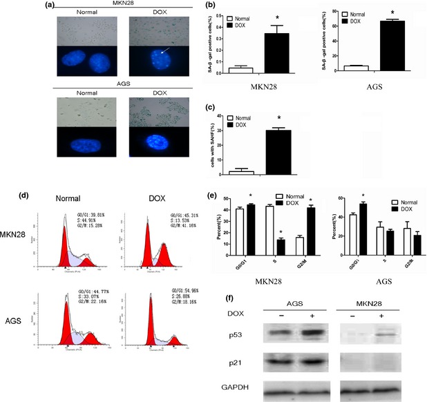
Figure 1.

Low concentration of doxorubicin (DOX) induced gastric cancer cells to senescence. Gastric cancer cells were treated with DOX for 2 h (MKN28: 25 μm, AGS: 0.1 μm). After 48 h of culture in drug‐free medium, cells were stained with SA‐β‐gal staining solution (pH 6.0) and DAPI, then cell images were captured. White arrow indicates senescence‐associated heterochromatin foci (a). The positive rate of SA‐β‐gal staining was increased in both cell lines (b). Percentage of senescence‐associated heterochromatin foci‐positive cells scored in normal and DOX group of MKN28 (c). Cell cycle was analysed by flow cytometry and cells arrested in G0/G1 phase were increased. Besides, there was also a significant increase in the proportion of cells in G2/M phase in MKN28 cells (d, e). A representative Western blot shows higher levels of p53 and p21 proteins in AGS, but only p53 increased in MKN28 after treatment of DOX (f). *Indicates a significant (P < 0.05) difference between normal and DOX group.
