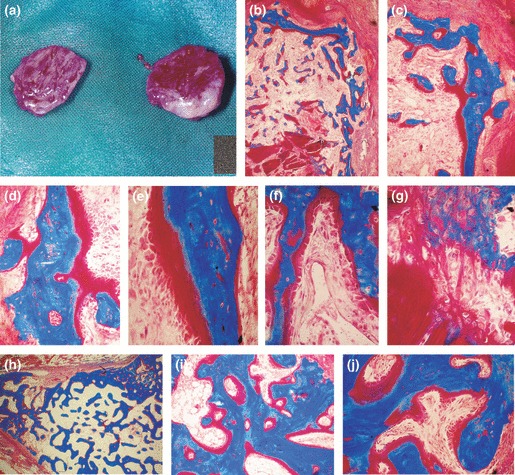
Figure 2.

Induction of bone formation by 5 μg recombinant hTGF‐β2 reconstituted with insoluble collagenous bone matrix and implanted in the rectus abdominis muscle of adult baboons Papio ursinus. (a) Induction of large corticalized ossicles by 5 μg hTGF‐β2 harvested on day 30. (b, c) Corticalized mineralized bone covered by osteoid seams induced by 5 μg hTGF‐β2 in the rectus abdominis muscle. (d–f) High power views showing mineralized bone in blue surfaced by large osteoid seams populated by contiguous osteoblasts facing invading capillaries (f). (g) Induction of bone formation within dissolving collagenous matrix as carrier, induction of osteoblastic‐like cells and mineralization in blue of the newly formed bone matrix. (h–j) Mineralized newly formed bone surfaced by osteoid seams 90 days after implantation of 5 μg hTGF‐β2. (j) Thick osteoid seams covered by contiguous osteoblasts. Undecalcified sections cut at 5 μm and stained free‐floating with Goldner’s trichrome. (b) original magnification ×25; (c) original magnification ×75 (d–g) original magnification ×125, 175, 125 and 175 respectively; (h–j) original magnification ×15, 75, 125 respectively.
