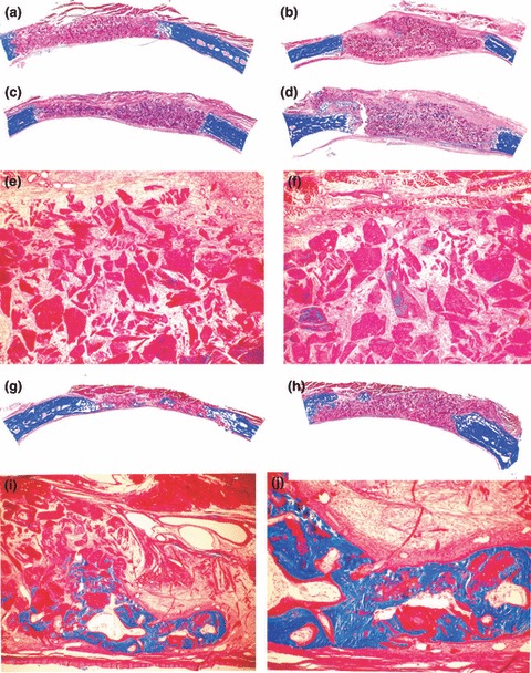
Figure 4.

Morphology of calvarial repair and induction of bone formation by hTGF‐β2 osteogenic devices without the addition of minced fragments of autogenous rectus abdominis muscle and harvested on day 30 and 90 after implantation. Lack of bone induction in calvarial defects harvested on day 30 after implantation of 100 (a, c) and 250 (b, d) μg hTGF‐β2. Note greater surface area of the implanted osteogenic devices in specimens of 250 μg hTGF‐β2 (b, d). (e, f) Higher power views of (c) and (d) showing scattered remnants of collagenous matrix as carrier with fibrovascular invasion but lack of bone differentiation. (g, h) Calvarial specimens harvested on day 90 after implantation of 100 (g) and 250 (h) μg hTGF‐β2 showing scattered islands of newly formed and mineralized bone in blue across the treated defects. (i, j) High power views of (g) showing mineralized bone in blue covered by osteoid seams and scattered remnants of the collagenous matrix as carrier. Undecalcified sections cut at 5 μm stained free‐floating with Goldner’s trichrome. (a, b, c, d, g, h) original magnification ×2.7; (e, f) original magnification ×37; (i, j) original magnification ×75 and 25 respectively.
