Figure 5.

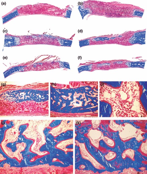
Morphology of calvarial regeneration and induction of bone formation in calvarial defects implanted with 100 and 250 μg hTGF‐β2 with the addition of morcellized fragments of autogenous rectus abdominis muscle and harvested on day 30 (a, b) and 90 (c–k) after implantation. (a, b) Lack of bone differentiation in calvarial defects implanted with 100 (a) and 250 (b) μg hTGF‐β2 showing minimal bone formation at the severed calvarial margins. (c, e, d, f) Reconstitution of the hTGF‐β2 osteogenic device with autogenous fragments of minced rectus abdominis muscle partially restores the biological activity of the hTGF‐β2 protein, resulting in the induction of large islands of mineralized bone on day 90 after implantation of 100 (c, e) and 250 μg (d, f) hTGF‐β2. (g–i) Higher power views of newly formed mineralized bone covered by osteoid seams with detail (i) of the induction of hematopoietic marrow on day 90. (j, k) Detail of mineralized trabecular bone in blue covered by osteoid seams populated by contiguous osteoblasts after the addition of morcellized fragments of autogenous rectus abdominis muscle on day 90. Undecalcified sections cut at 5 μm stained free‐floating with Goldner’s trichrome. (a–f) original magnification ×2.7; (g–i) original magnification ×25, 57, 175 respectively; (j, k) original magnification ×75.
