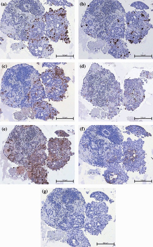
Figure 1.

Photomicrographs of hESC‐derived ILCs after 36 days of in vitro differentiation and immediately prior to transplantation. Serial sections stained by immunohistochemistry for: (a) insulin, (b) glucagon, (c) human C‐peptide, (d) pro‐hormone convertase 1/3, (e) pro‐hormone convertase 2, (f) cytokeratin‐19 and (g) representative isotype control. Magnification ×200; scale bar indicates 100 microns.
