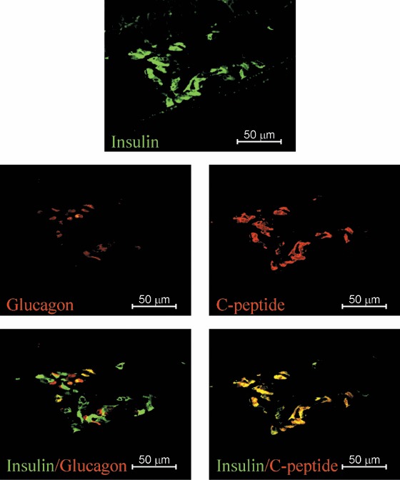
Figure 2.

Photomicrographs of a hESC‐derived ILC graft‐bearing kidney removed 1‐day post‐transplantation. Serial sections double stained with anti‐insulin (green) and antiglucagon (red) or double stained with antihuman‐C‐peptide (red) and anti‐insulin (green), specific antibodies as indicated. Magnification ×400; scale bar indicates 50 microns.
