
Figure 7.
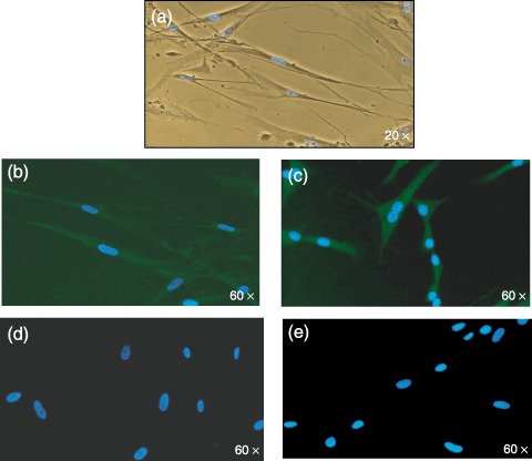
(a) Morphology of ADMSCs after exposure to endothelial conditioning medium (×20 magnification). Fluorescence image of ADMSCs after immunostaining with mouse–antihuman monoclonal antibodies for von Willebrand factor (b) and VE‐cadherin (c). (d) and (e) are the negative controls. Nuclei counterstained with DAPI (blue) (×60 magnification).