
Figure 5.
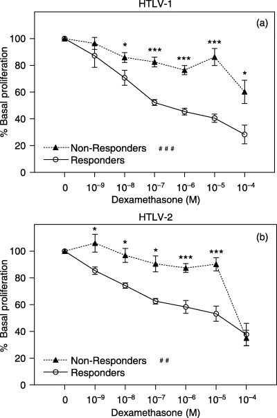
Peripheral T‐cell sensitivity to DEX in responders/non‐responders, HTLV‐infected patients. Glucocorticoid sensitivity was assessed by incubating PBMCs with PHA 1% and increasing concentrations of DEX for 96 h. Cell proliferation was estimated by MTT assay. OD was determined at wavelengths of 492 and 630 nm. Data are shown as percentage of base line cell proliferation (100% = PHA 1% without steroids). (a) HTLV‐I–infected subjects (responders: n = 10; non‐responders: n = 8); (b) HTLV‐II–infected subjects (responders: n = 6; non‐responders: n = 4). Statistical significance differences in T‐cell sensitivity to isolated DEX concentrations are indicated: *P < 0.05, **P < 0.01; ***P < 0.001. Statistical interaction of T‐cell sensitivity to variation of DEX concentrations between groups indicated: ##P < 0.01; ###P < 0.001.