
Figure 3.
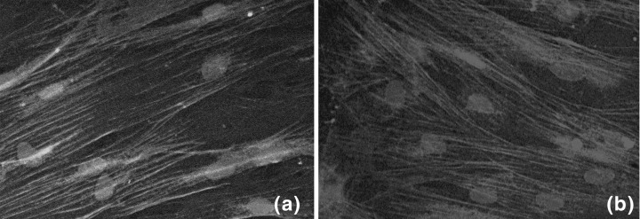
Data showing the phenotypic characterization of SMCs. (a) α‐SMA stained SMPC from the fourth passage; (b) calponin‐stained SMPC from the fourth passage and in both cases nuclei were developed with Sytox Blue. All images by confocal microscopy. Magnification, 60×.