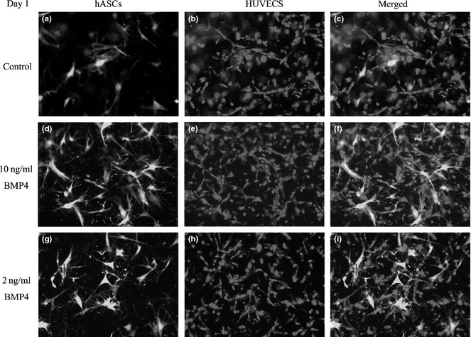
Figure 4.

Formation of 3D vascular network in vitro by day 1. In experimental group A, ASCs are labeled with GFP (a). HUVECs were cultured with hASCs. HUVECs began to proliferate and contact each other to form short tubule‐like structures (a, b). hASCs were found distributed around the HUVECs (c). In group B, more tubule‐like structures were observed (d–f). Group C was similar to group B (g–i).
