Figure 6.

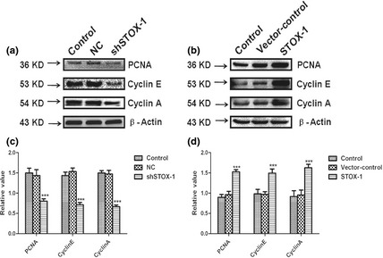
Expression of PCNA, cyclin E and cyclin A under normal culture conditions and after transfection with shSTOX1 or STOX1 over‐expression lentivirus. Protein levels of PCNA, cyclin E, cyclin A and β‐actin were analyzed by western‐blotting. (a, c) Densitometric quantification of protein bands showed significant reduction in PCNA, cyclin E, cyclin A after 48 h transfection with STOX1 shRNA lentivirus, whereas mismatch shRNA with only one nucleotide exchange (NC) showed no significant effect. (b, d) Densitometric quantification of protein bands showed significant increase in PCNA, cyclin E, cyclin A expression after 48 h transfection with STOX1 over‐expression lentivirus, whereas paired vector control showed no significant effect. Densitometric quantification was performed with at least three western‐blots from three different experiments. Epithelial cells transducted by over‐expression lentivirus, exhibited over 60% higher level of PCNA, cyclin E and cyclin A than the normal group, while in RNAi transfected cells, expression of PCNA, cyclin E and cyclin A was reduced by around 50%. Data are shown as means ± SD (n = 3, ***P < 0.001).
