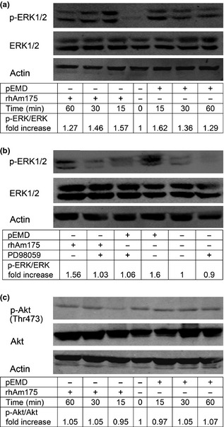
Figure 5.

Western blot analysis of total and phosphorylated ERK 1/2 and Akt/ PKB in human PDLF s after stimulation with rHhAm175 (10 μg/ml) or p EMD (200 μg/ml). Ratio between treatment group and negative control, obtained by densitometric analysis, is displayed under each lane. Experiments were performed in duplicate or triplicate and representative results are shown. (a) Quiescent cells were stimulated with either pEMD or rHhAm175 for 0–60 min. Lower bands showed abundance of total ERK as the loading control. (b) Quiescent cells (60‐min pre‐treatment of 20‐μm PD98059) were analysed 15 min after addition of rHhAm175 or pEMD to cells. Induced ERK phosphorylation could be abrogated by PD98059 and basal p‐ERK levels were not altered by the inhibitor. (c) Levels of total and phosphorylated Akt/PKB in human PDLFs at 0, 15, 30 or 60 min after stimulation with rHhAm175 or pEMD.
