
Figure 5.
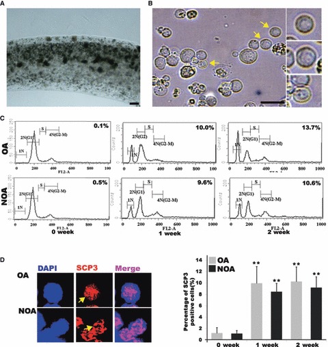
In vitro differentiation into sperm lineage cells. (A) Encapsulation with calcium alginate (B) Putative differentiated germ cells with diameter of 7–20 μm. Putative round spermatids (in small squares); yellow arrows indicate putative round spermatids (C) DNA content of in vitro differentiated SSCs from obstructive azoospermia (OA) and non‐obstructive azoospermia (NOA). 1N represents haploid germ cells, 2N represents diploid cells and 4N represents tetraploid cells. S‐phase represents synthesizing DNA. (D) SCP3 staining in putative meiotic germ cells from in vitro differentiated SSCs. Staining (left figure) and percentage (right figure) of Cy3‐SCP3‐stained cells during in vitro differentiation of SSCs. OA, obstructive azoospermic; NOA, non‐obstructive azoospermia; yellow arrows indicate putative meiotic germ cells. Bar = 50 μm. **Significantly different (P < 0.05).