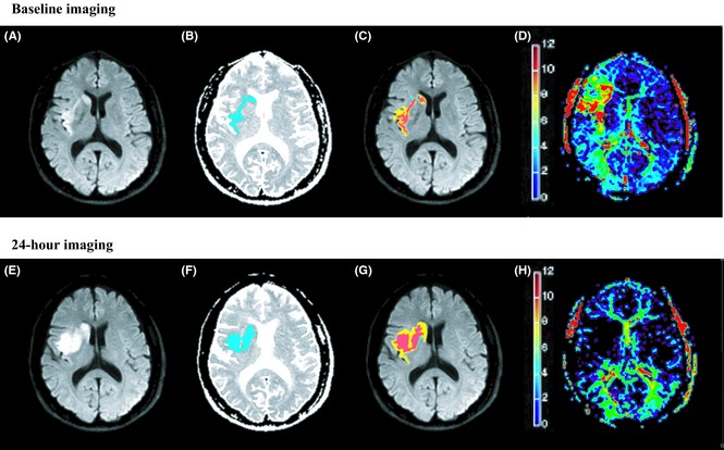
Figure 1.

A 56‐year‐old male obtained intravenous thrombolysis at 218 min after stroke onset. The DWI lesion (A and E. areas of visual hyperintensity) was coregistered with the hypointensity area on ADC map (B and F. blue areas). The baseline infarct (C. red areas indicate A overlap B) volume was 2 mL. Comparing with the baseline Tmax map (D. based on the color bar, signals covered from yellow to red indicated Tmax > 6 seconds tissues), 24‐h Tmax map (H.) confirmed that he achieved total reperfusion after thrombolysis. The final infarct (G. red areas) volume on 24‐h DWI was 9.2 mL, which proved the infarct progression.
