Figure 2.

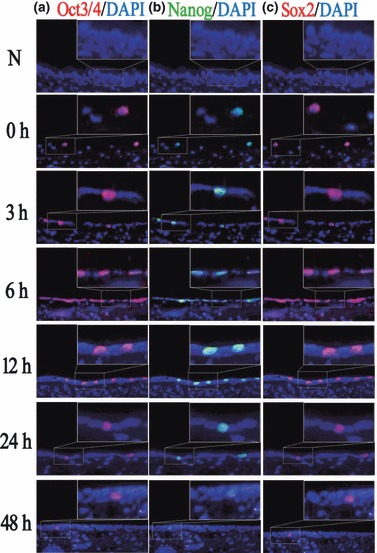
Immunofluorescence staining of Oct3/4, Nanog and Sox2 during recovery from injury induced by 5‐FU. (a) In untreated rat tracheal epithelium, Oct3/4 was not expressed. After treatment with 5‐FU, the number of Oct3/4‐positive cells increased gradually, reaching its peak at about 6 h, then decreased gradually and returned to its normal levels at about 48 h. (b,c) Expression tendency of Nanog and Sox2 was similar to that of Oct3/4, as they all are located in the same cells (original magnification ×400).
