
Figure 3.
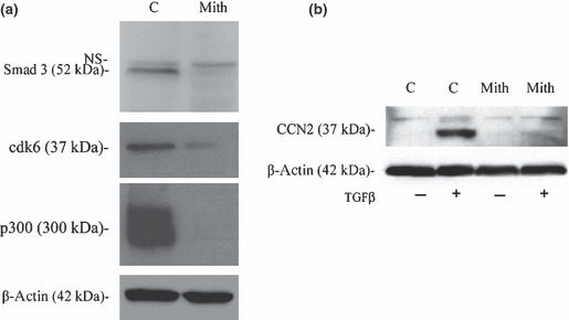
Mithramycin decreases protein levels in huma gingival fibroblasts. (a) After culturing the cells to 30–40% confluence, HGF were either treated with mithramycin (100 nm) or DMSO (mith or C) for 24 h. Cells were lysed and equal concentrations (25 μg) protein were subjected to SDS/PAGE and western blot analysis with anti‐Smad3, anti‐cdk6 and anti‐p300 antibodies. Blots were also probed with anti‐β‐actin antibodies show that lanes were loaded equally. (b) After culturing the cells to 30–40% confluence, HGF were cultured in DMEM containing 0.5% serum overnight, treated with mithramycin (100 nm) or DMSO for 1 h and TGFβ1 (4 ng/ml) for 24 h. Cells were lysed and equal concentrations (25 μg) of protein were subjected to SDS/PAGE and western blot analysis with anti‐CTGF and anti‐β‐actin antibodies.