
Figure 4.
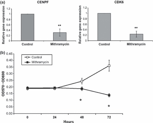
Mitramycin supressed proliferation of HGF (a) mRNA expression. RNA extracted using the Trizol kit from either mithramycin‐ (100 nm) or DMSO‐treated HGF was used in RT‐PCR analysis. Shown are the relative mRNA expression of CENPF and CDK6. Values are expressed as mean ± SD (n = 3). **P < 0.01 (b) proliferation assay. The effects of mithramycin (100 nm) on cell proliferation were assessed using the MTT assay. Cell number was determined using an ELISA plate reader at a wavelength of 570 nm. At 0 and 24 h, after treatment with mithramycin (100 nm), cell number is not significantly different (P > 0.05). There is a significant decrease in proliferation at 48 and 72 h after mithramycin treatment (P < 0.05). Similar results were obtained with human derma (foreskin) fibroblasts (not shown).