
Figure 5.
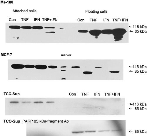
PARP‐cleavage in cells treated for 1 day with TNF, IFN, and TNF + IFN simultaneously. In apoptotic cells, the functional 116 kDa PARP molecule is degraded into a 85‐ and a 21‐kDa component. The antibody used in western blots of the three upper rows recognized the 116 and the 85 kDa proteins. Protein preparations of cells treated with the indicated cytokines were loaded into the respective lanes. The bottom row shows data obtained with an antibody against the 85 kDa cleaved fragment of PARP in TCC‐Sup cells. It is detected only in the floating cells.