Figure 4.

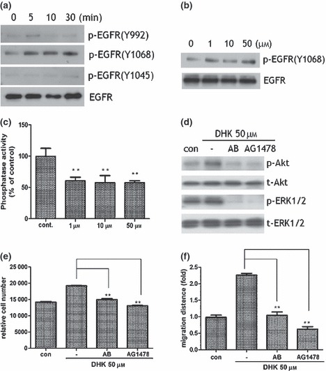
EGFR involvement in KSC stimulation. Note that DHK increased phosphorylation of EGFR. DHK significantly increased EGFR phosphorylation as shown in western blot analysis. Phosphorylation of Y1068‐EGFR increased just a few minutes after DHK treatment (a). Expression of Y1068‐phosphorylated EGFR increased in a dose‐dependent manner (b). DHK treatment significantly inhibited activity of protein tyrosine phosphotase activity in the membrane fraction of KSCs (c). EGFR neutralizing antibody (1 μg total protein) and chemical inhibitor (AG1478, 1 μm concentration) significantly attenuated phosphorylation of Akt and ERK1/2 signal pathways (d). In addition, proliferation (e) and migration (f) of KSCs were significantly reduced by antibody and inhibitor treatment. **P < 0.01.
