
Figure 1.
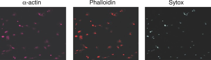
Immunohistochemical analysis of isolated cardiomyocytes. Cardiomyocytes were isolated from heart ventricles of newborn rats as described in the Materials and Methods section. They were plated in 0.1% serum medium and incubated for 2 days before staining with antibodies against sarcomeric α‐actin (actin), non‐specific staining of actin with phalloidin and DNA stained with Sytox Green nucleic acid.