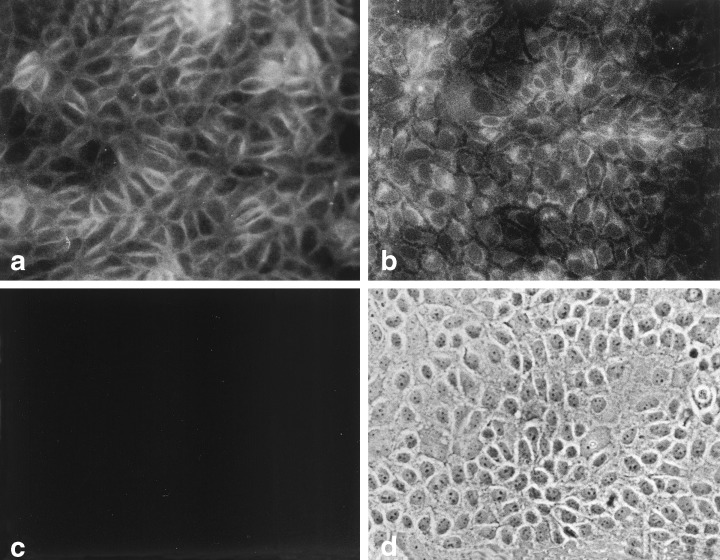
Figure 3.

Immunocytochemistry of LA7 cells for growth factors. (a) Fluorescent image of cells incubated with rabbit anti‐TGFα. (b) Fluorescent image of cells incubated with rabbit anti‐bFGF. (c) Fluorescent image of cells incubated with rabbit pre‐immune serum and the FITC‐conjugated second stage antibody. (d) Phase contrast image of cells in field (c). 200 ×.
