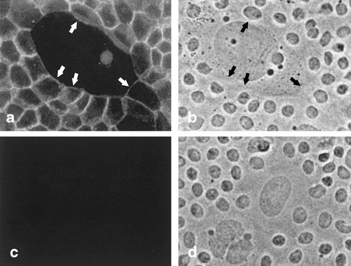
Figure 6.

Adherens junctions between MMEC and an irradiated feeder LA7 cell. (a) Fluorescent image of cells incubated with antibody to E‐cadherin. Adherens junctions line borders between MMEC on the edges of the field. Arrows in (a) and (b) point to sections of the junctions that line the border between MMEC and the large LA7 feeder cell in the centre of the field. (b) Phase contrast image of cells in (a). (c) Fluorescent image of a field of cells incubated with control rat serum. (d) Phase contrast image of cells in (c). 200 ×.
