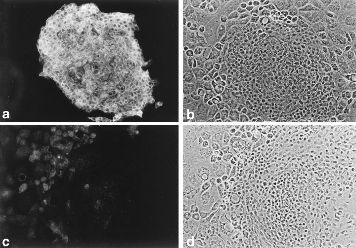
Figure 7.

Keratin 8 in MMEC. (a) Fluorescent image of an MMEC colony surrounded by dying feeder LA7 cells, after exposure to a rat anti‐keratin 8 serum and visualized with an FITC‐conjugated antibody to rat IgG. (b) Phase contrast image of field (a). (c) Fluorescent image of control cells exposed to pre‐immune rat serum. (d) Phase contrast view of field (c). An MMEC colony is on the right and feeder cells on the left. 100 ×.
