Figure 1.

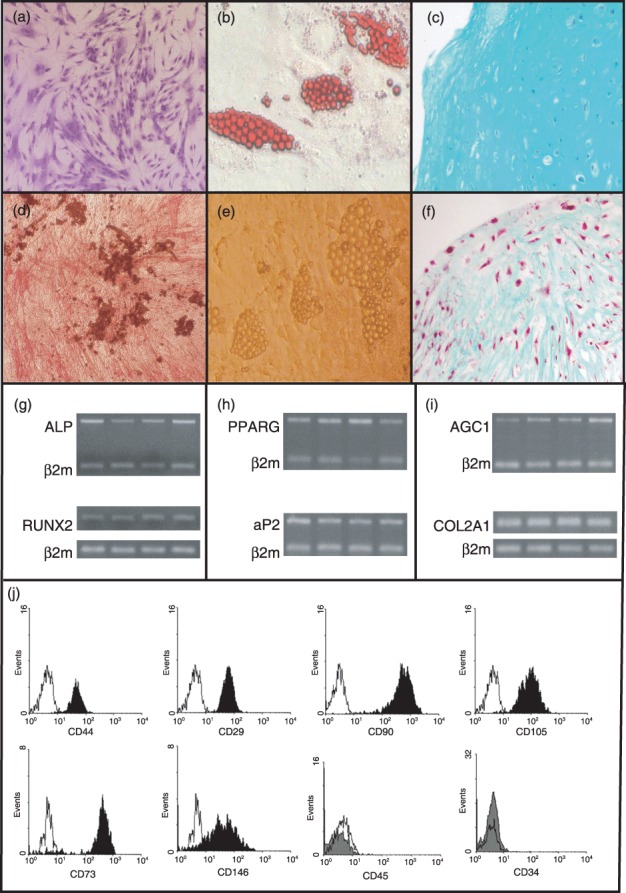
Differentiation potential and immunophenotypic characteristics of human mesenchymal stem cells (hMSCs). Culture expanded hMSCs from P2 stained with Giemsa, exhibiting characteristic spindle‐shaped morphology (a). Differentiated hMSCs from P2 towards osteogenic (d), adipogenic (b, e), and chondrogenic (c, f) lineages. Cell differentiation was identified with alkaline phosphatase (ALP)/Von Kossa (d), oil red O (b), Masson's (c) and alcian blue (f ) staining. Specific gene mRNA expression of P2 hMSCs upon differentiation towards the osteogenic (g), adipogenic (h), and chondrogenic (i) lineages. Lower panel ( j) shows representative plots from flow cytometric analysis of hMSCs at P2 stained with surface monoclonal antibodies. Black histogram bars indicate positive markers, and grey filled histogram bars depict the negative markers, in comparison to isotype‐matched controls (open histograms). Cells were also negative for CD14 (data not shown).
